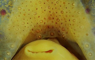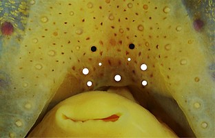


Figure. Ventral view of the funnel-groove region of W. scintillans, 37 mm ML, mature male. Top - Photograph of a preserved squid. Middle - In the photograph, white dots placed above the "white" photophores of the funnel groove. Black dots placed above the most posteromedial "red" photophores of the ventral head to mark the mark the anterolateral edges of the funnel groove. Bottom - This drawing shows the same dots seen in the picture above. Colors and connecting lines added to aid comparisons with other species. Images by R. Young.


 Go to quick links
Go to quick search
Go to navigation for this section of the ToL site
Go to detailed links for the ToL site