Click on an image to view larger version & data in a new window

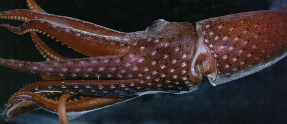
Figure. Ventral-oblique view of H. cerasina, Hawaiian waters. Photograph by R. young.
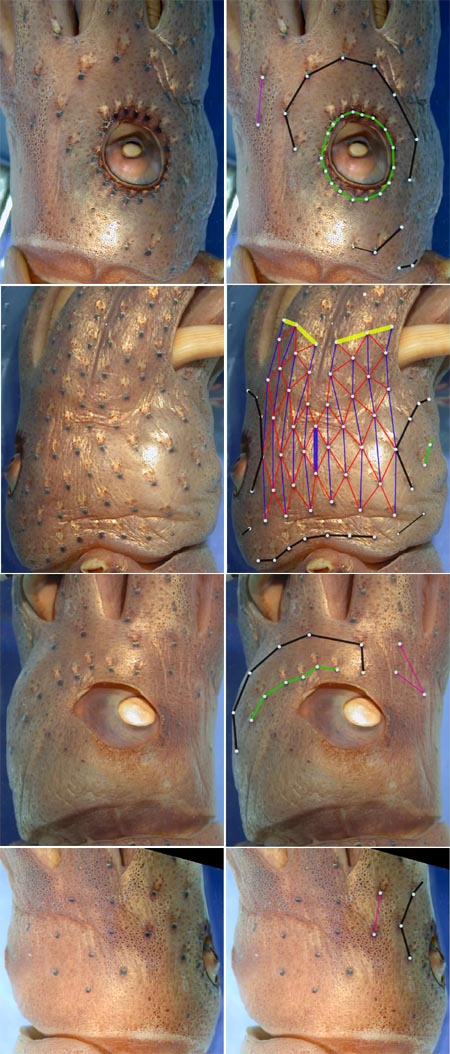
Photographs below are of a preserved squid and show the basic photophore groups, from different views, with superimposed lines and dots to emphasize the photophore groups. Photographs below left are the same pictures without superimposed lines and dots so that the photophores can be seen without obstruction. The superimposed lines are formed by first placing white dots over the pigmented, photogenic cups of all photophore in a particular group. The dots are then joined by a line of the appropriate color.
The color coding of the lines is as follows:
- Midline Series
- Ventral Matrix
- longitudinal series (thin blue lines)
- Basal Arm IV Row
- Right Eyelid Series
- Left Eyelid Series
- 2° Right Eyelid Series
- 2° Left Eyelid Series
- Basal row
- Right basal series
- Left basal series
- Right Accessory Series
- Left Accessory Series

Click on an image to view larger version & data in a new window

Figure. Various views of the head of H. cerasina, 60 mm ML, Hawaiian waters, preserved. Photographs by R. Young.

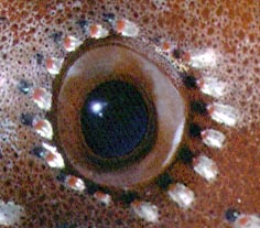
Figure. Lateral view of the photophores of the right eyelid in a living H. cerasina. Photograph by R. Young.
About This Page
Richard E. Young

University of Hawaii, Honolulu, HI, USA
Michael Vecchione

National Museum of Natural History, Washington, D. C. , USA
Page copyright © 2003
Richard E. Young
and
Michael Vecchione
 Page: Tree of Life
Histioteuthis cerasina: Photophore Pictures
Authored by
Richard E. Young and Michael Vecchione.
The TEXT of this page is licensed under the
Creative Commons Attribution-NonCommercial License - Version 3.0. Note that images and other media
featured on this page are each governed by their own license, and they may or may not be available
for reuse. Click on an image or a media link to access the media data window, which provides the
relevant licensing information. For the general terms and conditions of ToL material reuse and
redistribution, please see the Tree of Life Copyright
Policies.


 Go to quick links
Go to quick search
Go to navigation for this section of the ToL site
Go to detailed links for the ToL site