Three hooks of A. lichtensteini type B, were examined with a scanning electron microscope (SEM). Their relative sizes are seen in the photograph below. The 6th hook from the proximal end of the club in the ventral series (ie, hook V6) was the largest hook of all the club hooks. The hook situated two hooks more distally (hook V8) was also examined as was the hook from the dorsal series (hook D5) that lies opposite but slightly proximal to hook V6. Proceeding from left to right the hooks are D5, V8 and V6. Note the presence of a distinct "hairlip" on the hooks of the ventral series and the large size difference between hooks V6 and D5.

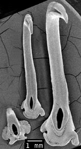
Figure. Frontal views of the three hooks (D5, left; V8, middle; V6,right) of A. lichtensteini examined, 157 mm ML, 05°30'S, 16°28'W. SEM photograph by R. Young.

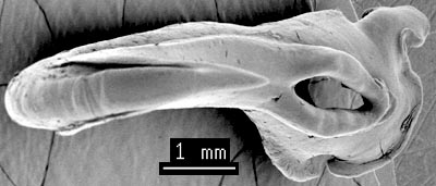
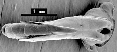
Figure. Top-frontal views of the two hooks of the ventral series. Left is the largest hook (V6) and right is hook V8. The lateral claw-grooves on the hook extend nearly to the hook tip. They do not join near the hook tip. The medial lateral lobe of V6 is greatly reduced and nearly absent in V8. SEM photograph by R. Young.

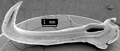

Figure. Oblique-side views of the two ventral-series hooks. Top - Hook V6. Bottom - Hook V8. SEM photograph by R. Young.

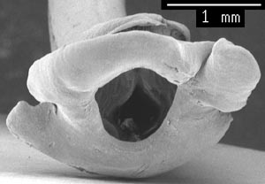
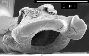
Figure. Oblique views of the attachment areas. Left - Hook V6. Right - Hook V8. The hairlip appears less accentuated in hook V8. SEM photograph by R. Young.

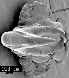
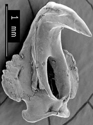
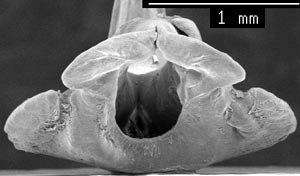
Figure. Various views of hook D5. Top, left - Top-frontal view. Top, right - Side-oblique view. Bottom - Attachment-site view. The lateral claw-grooves do not meet (same as the ventral series hooks). The hook is bilaterally symmetrical as is typical of onychoteuthid dorsal-series hooks. SEM photograph by R. Young.


 Go to quick links
Go to quick search
Go to navigation for this section of the ToL site
Go to detailed links for the ToL site