|
ID
|
Thumbnail |
Media Data |
| 30871 |

|
|
Specimen Condition
|
Live Specimen
|
|
Life Cycle Stage
|
Trophozoites
|
|
Copyright
|
©

|
|
Image Use
|
restricted
|
|
Attached to Group
|
Gregarina: view page image collection
|
|
Title
|
gregarine_lmcopy.jpg
|
|
Image Content
|
Specimen(s)
|
|
ID
|
30871
|
|
| 30872 |

|
|
Specimen Condition
|
Live Specimen
|
|
Copyright
|
©

|
|
Image Use
|
restricted
|
|
Attached to Group
|
Gregarina: view page image collection
|
|
Title
|
hosts.jpg
|
|
ID
|
30872
|
|
| 30873 |

|
|
Comments
|
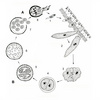
Illustration of the general life cycle of gregarines
|
|
Reference
|
modified after Vivier, E. and I. Desportes. 1990. Apicomplexa. In: Margulis, L., J.O. Corliss, M. Melkonian, and D.J. Chapman, eds. 1990. Handbook of the Protoctista; the structure, cultivation, habits and life histories of the eukaryotic microorganisms and their descendants exclusive of animals, plants and fungi. Jones and Bartlett Publishers. Boston. pp. 549-573.
|
|
Copyright
|
©

|
|
Image Use
|
restricted
|
|
Attached to Group
|
Gregarina: view page image collection
|
|
Title
|
general_lifecycle01copy.jpg
|
|
Image Type
|
Diagram
|
|
ID
|
30873
|
|
| 30874 |

|
|
Scientific Name
|
Monocystis agilis
|
|
Comments
|
An illustration of the Monocystis life cycle. The infection starts with the earthworm ingesting an oocyst (1, 2) that releases sporozoites (3, 4) within the intestines (green). The sporozoite is motile and penetrates the intestinal wall (5) and enters the dorsal blood vessel (6). The sporozoite enters the seminal vesicles (yellow) via the (dark red) dorsoventral hearts (7). The sporozoites feed on the developing spermatocytes (8) in the wall of the seminal vesicle. The gregarines then move into the lumen of the vesicle where they mature into trophozoites. Each ball of sperm within the seminal vesicle contains a young trophozoite (9). These trophozoites are covered by remnants of the sperm cells and often superficially take on the appearance of a ciliated eukaryotic cell. After consuming the sperm ball, the now mature trophozoites pair up in syzygy (10). The trophozoites develop into gamonts, and a gametocyst wall forms around each pair (11). Each of the two gamonts undergoes multiple nuclear divisions to produce many nuclei developing into gametes. Two gametes each fuse and form zygotes, which are surrounded by oocyst walls (12). Within this oocyst, the diploid zygote undergoes meiosis and then mitosis to produce eight haploid daughter cells by a process known as sporogony. The gametocysts, or oocysts if they have been released, leave the earthworm through the male genital pore and are liberated into the soil (13, 14). Infection of a new host occurs by oral ingestion of an oocyst (or perhaps through the process of mutual cross fertilization during host sexual reproduction).
|
|
Reference
|
modified after Olsen, O.W. 1974. Animal parasites: Their life cycles and ecology. Dover Publications, Inc., New York.
|
|
Copyright
|
©

|
|
Image Use
|
restricted
|
|
Attached to Group
|
Monocystids (Gregarina): view page image collection
|
|
Title
|
monocystis_lifecycle_newcopy.jpg
|
|
Image Type
|
Drawing/Painting
|
|
ID
|
30874
|
|
| 30933 |

|
|
| 31003 |

|
|
Scientific Name
|
Selenidium terebellae
|
|
Specimen Condition
|
Live Specimen
|
|
Life Cycle Stage
|
Trophozoite
|
|
Movie Use
|
 This media file is licensed under the Creative Commons Attribution-NonCommercial License - Version 3.0. This media file is licensed under the Creative Commons Attribution-NonCommercial License - Version 3.0.
|
|
Copyright
|
© Brian S. Leander

|
|
Attached to Group
|
Selenidium and relatives (Gregarina): view page movie collection
|
|
Title
|
Selenidium terebellae movement
|
|
Description
|
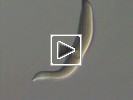
The movie shows the nematode-like bending, coiling and twisting movements of Selenidium terebellae from the intestines of a spaghetti worm, Thelepus.
|
|
Movie Type
|
Documentary
|
|
Movie Content
|
Specimen(s)
|
|
ID
|
31003
|
|
| 31004 |

|
|
| 31005 |

|
|
Scientific Name
|
Pterospora schizosoma
|
|
Specimen Condition
|
Live Specimen
|
|
Life Cycle Stage
|
Trophozoite
|
|
Movie Use
|
 This media file is licensed under the Creative Commons Attribution-NonCommercial License - Version 3.0. This media file is licensed under the Creative Commons Attribution-NonCommercial License - Version 3.0.
|
|
Copyright
|
© Brian S. Leander

|
|
Attached to Group
|
Urosporidae (Gregarina): view page movie collection
|
|
Title
|
Pterospora schizosoma dynamic peristalsis-like movements
|
|
Description
|
The movie shows the dynamic peristalsis-like movements of two attached trophozoites of Pterospora schizosoma, a urosporidian from the coelom of a bamboo worm, Axiothella.
|
|
Movie Type
|
Documentary
|
|
Movie Content
|
Specimen(s)
|
|
ID
|
31005
|
|
| 31006 |

|
|
Scientific Name
|
Selenidium vivax
|
|
Specimen Condition
|
Live Specimen
|
|
Life Cycle Stage
|
Trophozoite
|
|
Movie Use
|
 This media file is licensed under the Creative Commons Attribution-NonCommercial License - Version 3.0. This media file is licensed under the Creative Commons Attribution-NonCommercial License - Version 3.0.
|
|
Copyright
|
© Brian S. Leander

|
|
Attached to Group
|
Selenidium and relatives (Gregarina): view page movie collection
|
|
Title
|
Selenidium vivax movement
|
|
Description
|
The movie shows the bending and twisting movements of Selenidium vivax from the intestines of a peanut worm, Phascolosoma.
|
|
Movie Type
|
Documentary
|
|
Movie Content
|
Specimen(s)
|
|
ID
|
31006
|
|
| 31007 |

|
|
Scientific Name
|
Selenidium vivax
|
|
Specimen Condition
|
Live Specimen
|
|
Life Cycle Stage
|
Trophozoite
|
|
Movie Use
|
 This media file is licensed under the Creative Commons Attribution-NonCommercial License - Version 3.0. This media file is licensed under the Creative Commons Attribution-NonCommercial License - Version 3.0.
|
|
Copyright
|
© Brian S. Leander

|
|
Attached to Group
|
Selenidium and relatives (Gregarina): view page movie collection
|
|
Title
|
Selenidium vivax dynamic peristalsis-like movements
|
|
Description
|
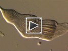
The movie shows bending movements and peristaltic waves passing from the posterior end the anterior end of the cell of Selenidium vivax from the intestines of a peanut worm, Phascolosoma. The flattened cell shape of the trophozoite and the active motility appear to be quite similar to that of detached proglottids of cestode flatworms.
|
|
Movie Type
|
Documentary
|
|
Movie Content
|
Specimen(s)
|
|
ID
|
31007
|
|


 This media file is licensed under the
  Go to quick links
Go to quick search
Go to navigation for this section of the ToL site
Go to detailed links for the ToL site