Go to 1° Unipartite Upper Beaks
Go to 3° Unipartite Upper Beaks
Go to Unipartite Upper-Beak Survey
SUBORDER SEPIOLIDA: 2 families
Phylogenetic relationships. At the base of the Decapodiformes there are five taxa (Idiosepiidae, Sepiidae, Spirulida, Sepiolida, Myopsida (= Loliginidae plus the monotypic Australiteuthidae). The Sepiolida is placed with the Sepiidae in the Tree of Life based on morphology but there is considerable uncertainty regarding relationships based on morphology among all five taxa. Using molecular data, Lindgren et al. (2012) with an extensive data set was unable to resolve relationships among them. Lindgren in (1910), using a Bayesian analysis, found the same 5 taxa basal to the Decapodiformes but within this clade found Sepiolida as sister to the Sepiidae with strong support but her Likelihood analysis found Sepiidae sister to Idiosepiidae with some support.
SEPIADARIIDAE: 2 genera
UPPER BEAK CHARACTERISTICS - Step: Absent.
- Shoulder Blade: Rather thick; lateral (=outer) surface of Shoulder Blade without thin layer of hyaline material.
- Shoulder Blade: Consists of two components (Inner and Outer). Components separated by distinct line. Inner component may form in situ.
- Hyaline Matrix: Forms the Middle Part of the Shoulder between the Shoulder Blade and the Palatoshoulder Ridge.
- Palatoshoulder Ridge. Apparently present but reduced, limited to the dorsal region of the Shoulder.
- Pigmentation of posterior surface of Hyaline Matrix: Mechanism unknown.
- Yellow line: Absent. A "White Patch" on oral surface near each jaw angle (a possible Yellow Line derivative (see: Doryteuthis, Loliginidae)), present.
Phylogenetic relationships.The Sepiadariidae have long been considered to be the earliest offshoot of the Sepiolida lineage (Naef 1921-23). This position has been supported by the molecular work of Lindgren, et al., 2012 and Pankey, et al., 2014.
Sepiadariidae: Structure
- EXTERNAL FEATURES
One beak of Sepiadarium sp. and two of Sepioloidea pacifica have been examined.
- INTERNAL STRUCTURE: Longitudinal cuts through the Shoulder
 Click on an image to view larger version & data in a new window
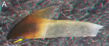
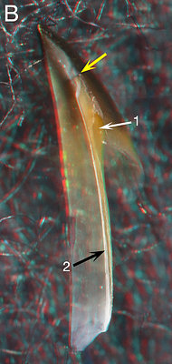
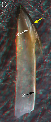


Figure. A-D - Sepiadarium sp., female, 22 mm ML (same beak as above). A: Side view showing the position of the cut. B - Oral-lateral view of cut beak. C - Oral view of cut beak. D - Photomicrograph (2D), oral view taken perpendicular to the cut surface. E - Sepioloidea pacifica, immature male, 15 mm ML, 0.5 mm ML. Photomicrograph (2D), oral view taken perpendicular to the cut surface.
ARROWS: 1 - Pigmented posteriior surface of the Hyaline Matrix. 2 - Hyaline material of the Lateral Wall. 3 - Palatoshoulder Ridge. D4 - Inner Component of the Shoulder Blade. E4 - Possible start of the Inner Component. 5 - Hyaline Matrix. 6 - Bar indicates width of the pigmented part of the Lateral Wall. Yellow arrows - Point to same spot on all beaks (anterioventrall end of the Shoulder Blade where cut was made).
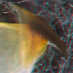 | 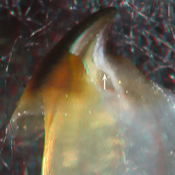 | 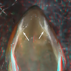 |
| Sepiadarium sp., female, 22 mm ML, Taiwanese waters. LEFT - Side view. MIDDLE - Oblique view. RIGHT - Oral-front view. | ||
The Shoulder has no Step, no Yellow Line and a slightly acute Jaw Angle (Left, Middle). A reduced Palatoshoulder Ridge (Middle, arrow, Right, arrows), however, appears to be present as does a White Patch (Middle, Right).
Interpretation of the structure of the Sepiadarium beak is problematical since the Hyaline Matrix is in the process of being pigmented. That is, the apparent Palatoshoulder Ridge, in this case, could be a manifestation of the final pigmentation of the Shoulder. Also, in the related Sepioloidea pacifica a 19 mm immature female, like the immature male (15 mm ML) seen above, shows little indication of a Palatoshoulder Ridge even though the Lateral Wall is about 3/4 pigmented.
SEPIOLIDAE: 4 subfamilies, 17 genera
UPPER BEAK CHARACTERISTICS
- Step: Absent.
- Shoulder Blade: Rather thick; lateral (=outer) surface of Shoulder Blade without thin layer of hyaline material.
- Shoulder Blade: Consists of two components (Inner and Outer). Components separated by distinct line.
- Hyaline Matrix: Apparently forms Middle Part of Shoulder in Rossinae and Heteroteuthinae (verification needed in both taxa); forms Inner (= medial) Part of Shoulder in Sepiolinae.
- Palatoshoulder Ridge: Varies from absent to remnant.
- Pigmentation of posterior surface of Hyaline Matrix: Inner Component of Shoulder Blade develops virtually simultaneously with the initial posterior pigment of the Lateral Wall (Euprymna).
- Yellow line: Absent. A "White Patch" on oral surface near each jaw angle, present but may be lost in some adults.
Sepiolidae: Structure
We examined mostly mature members of three subfamilies and, superficially, they are all quite similar. Unfortunately, critical growth stages were, generally, not available to cut. As a result considerably uncertainty exists on the shoulder structure.
- EXTERNAL FEATURES
One beak from a mature female and one from a very young female (?) was examined. The latter was too young to provide useful information and the former nearly too old.
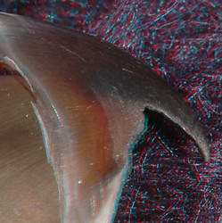
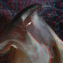
Rossia pacifica, mature female, 38 mm ML, 1.7 mm URL. LEFT - Side view. RIGHT -
Oblique view.The Shoulder has no Step, no Yellow Line and a slightly damaged but acute Jaw Angle (Left). A possible but reduced/remnant of a Palatoshoulder Ridge is present (Right, arrow) as is a White Patch (Right).
- INTERNAL STRUCTURE: Longitudinal cuts through the Shoulder
 Click on an image to view larger version & data in a new window
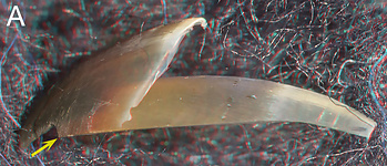
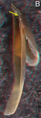
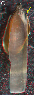


Figure. Rossia pacifica, mature female, 38 mm ML, 1l7 mm URL (same beak as above). A - Side view of beak fragment showing position of cut. B - Oral-lateral view of cut beak. C - Oral view of cut beak. D - Photomicrograph (2D), oral view taken perpendicular to cut surface. E - Same image as D but lower magnification.
ARROWS: 1 - Hyaline Matrix. 2 - Line that separates the two Components of the Shoulder Blade. 3 - Approximate point of Point of changeover (judging by thickness) from Hyaline Matrix to hyaline material. 4 - Hyaline material.
A White Patch is present (C) and a low ridge, a possible Palatoshoulder Ridge, medial to the Patch doesn't reach the cut surface; the darkened area lateral to the "ridge" suggests that the white area has little depth indicating that little remains of a Ridge at this stage. The Hyaline Matrix (1) is not very thick but its residue seem fairly extensive (3). Cuts through a younger beak are needed.
HETEROTEUTHINAE: 6 genera
- EXTERNAL FEATURES
We examined one beak of a mature male Iridoteuthis sp. A (below) and several Heteroteuthis hawaiiensis but the latter were not of a critical size and yielded no useful information.
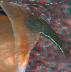
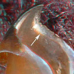
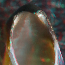
Iridoteuthis sp. A, mature male, 27 mm ML, off New Caledonia. LEFT - Side view. MIDDLE - Oblique view. RIGHT - Front view.
The Shoulder has no Step, no Yellow Line and a slightly acute Jaw Angle (Left). A remnant of a Palatoshoulder Ridge (note the lighter color) is present (Middle, arrow, Right) as is a White Patch (Middle, Right).
- INTERNAL STRUCTURE: Longitudinal cuts through the Shoulder
 Click on an image to view larger version & data in a new window
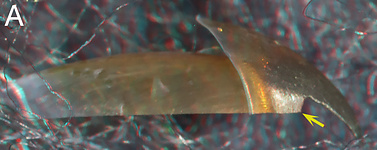

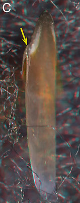

Figure. Iridoteuthis sp. A, mature male, 27 mm ML, 0.8 mm URL. A: Side view showing the position of the cut. Bt - Oral-lateral view of cut beak. C - Oral view of cut beak. D - Photomicrograph (2D), oral view perpendicular to the cut of the Shoulder.
ARROWS: 1 - Line that separates the two Components of the Shoulder Blade. 2 - Probable remnant of the Palatoshoulder Ridge (but see below). Yellow arrows - Point to same spot on all beaks (anterior end of the Shoulder Blade where cut was made).
D, 2 (above) appears be a remnant of the Palatoshoulder Ridge, that lacks well-defined edges, but could be the initial pigmentation of the Hyaline Matrix.
SEPIOLINAE: 5 genera
- EXTERNAL FEATURES
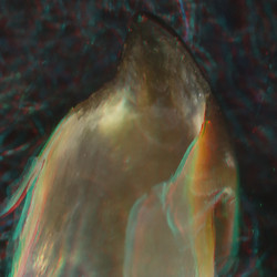
We have examined seven Euprymna ranging from 10-26 mm ML. This is the only sepiolid for which we had reasonable size range of beaks to study.
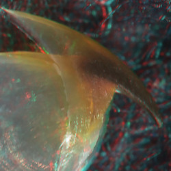
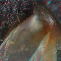
Euprymna scolopes, male, immature ?, 21 mm ML, 0.9 mm URL. LEFT - Side view. MIDDLE
- Oblique view.
Euprymna scolopes, mature female, 26 mm
ML, 1.3 mm URL. RIGHT - Oblique view.The Shoulder has no Step, no Yellow Line and a very slightly acute Jaw Angle (Left). A Palatoshoulder Ridge is absent (Middle, Right); a White Patch (Middle, Right) is present.
- INTERNAL STRUCTURE: Longitudinal cuts through the Shoulder
 Click on an image to view larger version & data in a new window
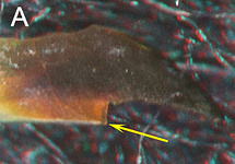
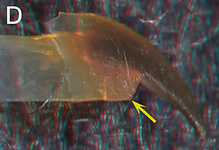
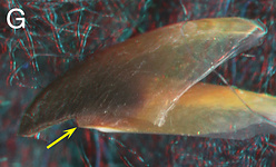

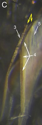

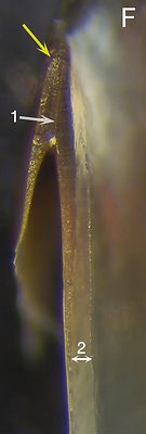

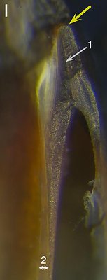
Figure. Euprymna scolopes. A-C: Male, immature, 15 mm ML, 0.8 mm URL. A - Side view showing the position of the cut. B - Oral view of the beak fragment. C - Photomicrograph (2D), oral view taken perpendicular to the cut of the Shoulder. D-F: Male, immature ?, 21 mm ML, 0.9 mm URL. D: Side view showing the position of the cut. E - Oral view of cut beak. F - Photomicrograph (2D), oral view taken perpendicular to the cut of the Shoulder. G-I: Mature female, 26 mm ML, 1.3 mm URL. G: Side view showing the position of the cut. H - Oral view of cut beak. I - Photomicrograph (2D), oral view taken perpendicular to the cut of the Shoulder.
ARROWS: 1 - Line that separates the two Components of the Shoulder Blade. 2 - Indicates width of hyaline material. 3 - Outer Component of the Shoulder Blade. 4 - Developing Inner Component of the Shoulder Blade. 5 - Outer edge of the Hyaline Matrix. Yellow arrows - Point to same spot on all beaks. Double arrows in Fig. C indicate a fracture at the tip of the Outer Component resulting from the cut.
The immature male has a Shoulder (C) that appears to be in the process of developing the Inner Component of the Shoulder Blade (enlarge this figure for details - i.e., click). This indicates that the Inner Component develops virtually simultaneously with the initial posterior pigment of the Lateral Wall. That is, there is no pigment gap between the two. The Shoulder of the larger male (F) shows a fully developed Inner Component and a reduction of the Hyaline Matrix. The large female has a much more robust Shoulder Blade (I), and a further reduced Hyaline Matrix. Due to the position of the cut close to the Jaw Angle, the cut passed through the White Patch.
ORDER UNCERTAIN, Superfamily - Bathyteuthoidea:
CHTENOPTERIGIDAE: 3+ species
UPPER BEAK CHARACTERISTICS
- Step:The largest Chtenopteryx (1.2 mm URL) that we have seen has no step in the shoulder.
- Shoulder Blade:Thick; without thin outer (= Lateral) layer of hyaline material.
- Shoulder Blade: Consists of two Components (Inner and Outer) separated by a distinct line.
- Hyaline Matrix: Forms Inner (= medial) Part of Shoulder.
- Palatoshoulder Ridge: Absent.
- Pigmentation of posterior surface of Hyaline Matrix: Mechanism unknown.
- Yellow line: Absent. White Patch absent.
Phylogenetic Relationships. On the basis of morphology, the ToL has placed the Bathyteuthidae with the Chtenopterygidae in the Bathyteuthoidea. The molecular work of Lindgren (2010) and Lindgren et. al. (2012) strongly supports the monophyly of the Bathyteuthoidea.
J.Z. Young (1991) found some important morphological similarities between Chtenopteryx and the Loliginidae (≅ Myopsida). R. Young, et al. (1998) on the basis of morphology placed the Loliginidae somewhere between the Oegopsida and the Sepioidea. Lindgren et al. (2012) placed the Bathyteuthoidea as basal to the Oegopsida with good support and placed the Loliginidae within the sepioid taxa, near the Bathyteuthoidea but without support.
The other member of the Bathyteuthoidea, the Bathyteuthidae, has a Type 2 Bipartite beak with some unresolved features. Presumably the 2° Unipartite beak of the Chtenopterygidae is due to convergent evolution. However, knowing that the Loliginidae could be a close relative, opens some interesting possibilities.
Chtenopterygidae: Structure
- EXTERNAL FEATURES
We examined six beaks of Chtenopteryx, involving more than one species, and cut two beaks.
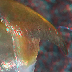
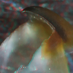
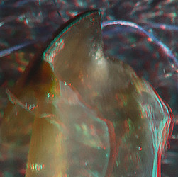
Chtenopteryx sp., male ?, immature, 35 mm ML, 0.8 mm URL. LEFT - Side view. MIDDLE -
Oblique view.Chtenopteryx sp., mature male, 32 mm ML,
0.7 mm URL. RIGHT - Oblique view.
The Shoulder has no Step, no Yellow Line and a Jaw Angle that is more of a curve than an angle (Left). A Palatoshoulder Ridge is absent (Middle, Right); a White Patch (Middle, Right) is absent.
- INTERNAL STRUCTURE: Longitudinal cuts through the Shoulder
 Click on an image to view larger version & data in a new window
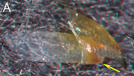
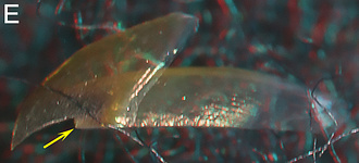
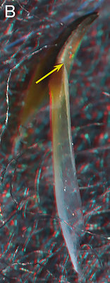

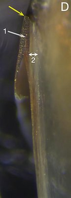

Figure. Chtenopteryx sp. A-D: Male ?, immature, 35 mm ML, 0.8 mm URL, with longitudinal cut through the Shoulder. A - Side view showing where the cut was made. B - Lateral-oral view of the cut beak. C - Oral view of the cut beak. D - Photomicrograph (2D), oral view taken perpendicular to the plane of the cut. Chtenopteryx sp. E-F: Mature male, 32 mm ML, 0.7 mm URL, with longitudinal cut through the shoulder. E - Side view showing where the cut was made. F - Photomicrograph (2D), oral view taken perpendicular to the plane of the cut.
ARROWS: 1 - Line that separates the two components of the Shoulder Blade. 2 - Indicates width of Hyaline Matrix. Yellow arrows - Point to same spot on all beaks (anterior end of the Shoulder Blade where cut was made).
The immature Chtenopteryx sp. has a fully developed double Shoulder Blade and a thick layer of Hyaline Matrix medial to the Shoulder blade. No Palatoshoulder Ridge is present. The lateral surface of the Shoulder Blade is bare (ie, no outer layer). The mature female has the same features.


 Go to quick links
Go to quick search
Go to navigation for this section of the ToL site
Go to detailed links for the ToL site