Sellaphora
David G. Mann


This tree diagram shows the relationships between several groups of organisms.
The root of the current tree connects the organisms featured in this tree to their containing group and the rest of the Tree of Life. The basal branching point in the tree represents the ancestor of the other groups in the tree. This ancestor diversified over time into several descendent subgroups, which are represented as internal nodes and terminal taxa to the right.

You can click on the root to travel down the Tree of Life all the way to the root of all Life, and you can click on the names of descendent subgroups to travel up the Tree of Life all the way to individual species.
For more information on ToL tree formatting, please see Interpreting the Tree or Classification. To learn more about phylogenetic trees, please visit our Phylogenetic Biology pages.
close boxNote: this tree is still under construction. It does not yet contain all known Sellaphora subgroups.
Many further Sellaphora species exist (both described and undescribed) but have not yet been added to the treeIntroduction
Species of Sellaphora have been known since c. 1840 but were classified in the genus Navicula for most of time up to 1990. During most of the 19th and 20th centuries, diatom taxonomy was based almost entirely on the structure of the frustule (the silicified cell wall of diatoms); other features, such as chloroplast morphology, reproductive characteristics or nuclear behaviour, were not considered. There is nothing particularly unusual about the structure of the frustule in Sellaphora and so the original classification of Sellaphora species in Navicula (then a catch-all for undistinguished boat-shaped diatoms) is understandable. However, if chloroplast morphology is taken into account, differences between Sellaphora and Navicula species are immediately apparent. This was first noticed by Mereschkowsky (1902), who gave the following description:
‘Valve small, symmetrical, linear to elliptical, with obtuse ends, terminal nodules distant; striae usually fine, connecting-zone simple. Endochrome composed of one plate, resting with its narrow median part on the surface of one of the valves, with four long prolongations along the connecting zones. Pyrenoid absent. A few elaeoplasts, sometimes represented by two libroplasts.’
The key feature was the shape of the chloroplast (Mereschkowsky’s ‘endochrome’), which is a single H-shaped plate; this is what gives the genus its name (Sellaphora = ‘saddle-bearer’). Most 20th century floras reverted to the 19th century practice of including Sellaphora in Navicula and the species now assigned to Sellaphora (see below) were not always put in the same group within Navicula. However, following SEM studies of the frustule and new observations of the living cell and sexual reproduction, Sellaphora was reinstated as a separate genus (Mann 1989, Round et al. 1990). Molecular genetic data have thus far supported the monophyly of Sellaphora.
The history and taxonomy of Sellaphora are discussed by Mann (1989), Mann et al. (2008) and Evans et al. (2008). The genus has been developed as a model system for studying and documenting speciation in freshwater diatoms, including DNA barcoding (Evans et al. 2007). More is known about the mating systems of Sellaphora species than in any other diatom genus (e.g. Mann 1999, Mann et al. 1999).
Characteristics
- freshwater epipelic diatoms
- plastid single, H-shaped, lying with its centre against the epivalve.
- pyrenoid ± tetrahedral during interphase, invaginated; one per plastid.
- plastid translocated onto the girdle before mitosis.
- division of plastid beginning before cytokinesis and before the previous division is complete, proceeding asymmetrically, and arresting before completion to produce the H-shape of interphase.
- 90º rotation of the plastid onto the epivalve occurring at the end of valve formation.
- frustule generally wider whan deep and usually lying in valve view
- valve outline elliptical, linear to lanceolate, with rounded, rostrate or capitate poles.
- striae uniseriate (rarely biseriate), not interrupted by lyre-shaped plain areas
- poroids small, round, containing hymenes.
- a narrow plain conopeum sometimes present on either side of the valve, extending out from the raphe-sternum; conopea sometimes continuous from pole to pole, elsewhere only from pole to centre.
- internal central raphe endings deflected towards the primary side of the valve.
- gametangia not surrounded by an obvious mucilage sheath; closely associated throughout meiosis and plasmogamy; opening only partially to allow gamete migration via a copulation aperture.
- one functional gamete and one apochlorotic cell formed per gametangium.
- gametes physiologically anisogamous: auxospore formed within one gametangium and expanding parallel to its apical axis.
References
Evans, K.M., Wortley, A.H. & Mann, D.G. (2007). An assessment of potential diatom “barcode” genes (cox1, rbcL, 18S and ITS rDNA) and their effectiveness in determining relationships in Sellaphora (Bacillariophyta). Protist 158: 349–364.
Evans, K.M., Wortley, A.H., Simpson, G.E., Chepurnov, V.A. & Mann, D.G. (2008). A molecular systematic approach to explore diversity within the Sellaphora pupula species complex (Bacillariophyta). Journal of Phycology 44: 215–231.
Mann, D.G. (1989). The diatom genus Sellaphora: separation from Navicula. British Phycological Journal 24: 1–20.
Mann, D.G. (1999). The species concept in diatoms (Phycological Reviews 18). Phycologia 38: 437–495.
Mann, D.G., Thomas, S.J. & Evans, K.M. (2008). Revision of the diatom genus Sellaphora: a first account of the larger species in the British Isles. Fottea 8: 15–78.
Mereschkowsky, C. (1902). On Sellaphora, a new genus of diatoms. Annals and Magazine of Natural History, series 7, 9: 185–195.
Round, F.E., Crawford, R.M. & Mann, D.G. (1990). The diatoms. Biology and morphology of the genera. Cambridge University Press, Cambridge, 747 pp.
Title Illustrations

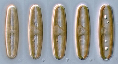
| Scientific Name | Sellaphora 'urban elliptical' deme |
|---|---|
| Specimen Condition | Live Specimen |
| Identified By | David G. Mann |
| Life Cycle Stage | vegetative phase |
| Body Part | single cell, focus series |
| View | valve view |
| Image Use |
 This media file is licensed under the Creative Commons Attribution-NonCommercial License - Version 3.0. This media file is licensed under the Creative Commons Attribution-NonCommercial License - Version 3.0.
|
| Copyright |
© 2008 David G. Mann

|
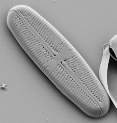
| Scientific Name | Sellaphora |
|---|---|
| Creator | Frieda Christie |
| Acknowledgements | This micrograph was taken by Frieda Christie, Royal Botanic Garden Edinburgh |
| Specimen Condition | Dead Specimen |
| Identified By | David G. Mann |
| Life Cycle Stage | Vegetative phase |
| Body Part | valve |
| View | exterior, SEM |
| Image Use |
 This media file is licensed under the Creative Commons Attribution-NonCommercial License - Version 3.0. This media file is licensed under the Creative Commons Attribution-NonCommercial License - Version 3.0.
|
| Copyright |
© 2008 David G. Mann

|
About This Page
This page is being developed as part of the Tree of Life Web Project Protist Diversity Workshop, co-sponsored by the Canadian Institute for Advanced Research (CIFAR) program in Integrated Microbial Biodiversity and the Tula Foundation.
David G. Mann

Royal Botanic Garden Edinburgh, United Kingdom
Correspondence regarding this page should be directed to David G. Mann at
d.mann@rbge.org.uk
Page copyright © 2008 David G. Mann
 Page: Tree of Life
Sellaphora .
Authored by
David G. Mann.
The TEXT of this page is licensed under the
Creative Commons Attribution-NonCommercial License - Version 3.0. Note that images and other media
featured on this page are each governed by their own license, and they may or may not be available
for reuse. Click on an image or a media link to access the media data window, which provides the
relevant licensing information. For the general terms and conditions of ToL material reuse and
redistribution, please see the Tree of Life Copyright
Policies.
- First online 07 February 2010
Citing this page:
Mann, David G. 2010. Sellaphora . Version 07 February 2010 (under construction). http://tolweb.org/Sellaphora/125733/2010.02.07 in The Tree of Life Web Project, http://tolweb.org/


 Go to quick links
Go to quick search
Go to navigation for this section of the ToL site
Go to detailed links for the ToL site