Leishmania
Julius Lukes- Leishmania donovani
- Leishmania infantum
- Leishmania major
- Leishmania tropica
- Leishmania mexicana
- Leishmania tarentolae
- Leishmania braziliensis
Note: this taxon list is still under construction. It does not yet contain all known Leishmania subgroups.
Introduction
The genus Leishmania contains several human pathogens. These parasites circulate among both cold- and warm-blooded vertebrates, being transmitted by dipterid phlebotomine flies of the genera Phlebotomus and Lutzomyia in the Old and New World, respectively (Dujardin 2006). However, unlike trypanosomes, they undergo dramatic morphological changes during their life cycle. In the vertebrates they exist as aflagellated, round amastigote stages invading mostly macrophages, while in the insect host flagellated promastigotes are found.
The life cycle commences with macrophages infected with amastigotes entering the vector during blood feeding. In the insect, the parasite multiplies in the form of promastigotes that progressively move into the mesenteron and are ready to infect the new host during another blood feeding. After entering the skin of the new host, promastigotes are quickly phagocytosed by host macrophages, where they undergo almost instant transformation into the amastigote stage. The compartment in which amastigotes reside in the macrophage, the so-called phagosome, eventually fuses with lysosomes creating a phagolysosome. While other pathogens are killed by the extremely low pH in this compartment, Leishmania cells proliferate there.
The genus Leishmania comprises dozens of species, many of which are known to infect humans. Individual Leishmania species cannot be distinguished on a morphological basis but only by biochemical and molecular approaches, such as isoenzyme typing, restriction fragment length polymorphism, species-specific PCR amplification and others (Schonian et al., 2003). Since there are dramatic differences in symptoms and clinical outcomes of the diseases caused by different Leishmania species, species diagnosis is critical for proper treatment. Collectively, the diseases are termed leishmaniasis; they can be divided into three main forms:
- cutaneous leishmaniasis is found in Southern Europe, North Africa, the Middle East and central Asia. Slightly different forms, named “dry” and “wet” ulcer (boil) are caused by L. tropica and L. major, respectively, as well as by several other less frequent species, some of them with dubious species status; L. mexicana is responsible for another cutaneous form of the disease found in Central America;
- mucocuteneous leishmaniasis occurs in Central and South America, where it is caused by a group of species and sub-species, mainly L. braziliensis. The most serious form of the disease, called espundia, can be fatal.
- visceral leishmaniasis – by far the most deadly form, distributed originally in the Mediterranean, where it is caused by L. infantum, was spread to South America with the European settlers. In this new locale it was called L. chagasi, but it was recently synonymized for good reasons with L. infantum (Lukes et al., 2007). Even more serious is the infection caused by L. donovani, encountered mostly in India, Bangladesh, Pakistan and Sudan, called cala-azar or black disease. It is an antroponosis (India) or zoonosis (East Africa) leading to massive hepato and/or splenomegaly, which becomes, if untreated, highly lethal usually within several months. Currently, it is being treated with drugs on the basis of pentamidin and amfotericin B.
References
Dujardin J.C. 2006. Risk factors in the spread of leishmanises: towards integrated monitoring? Trends Parasitol. 22: 4-6.
Schonian G., A. Nasereddin, N. Dinse, C. Schweynoch, H.D.F.H Schallig, W. Presber and C.L. Jaffe. 2003. PCR diagnosis and characterization of Leishmania in local and imported clinical samples. Diagn. Microbiol. Infect. Dis. 47: 349-358.
Title Illustrations

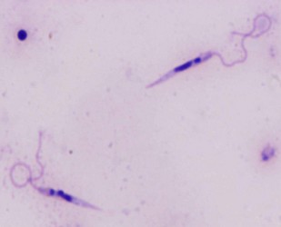
| Scientific Name | Leishmania major |
|---|---|
| Comments | from Promastigote (culture form) |
| Specimen Condition | Live Specimen |
| Copyright |
© 2008 Jan Votýpka

|
About This Page
This page is being developed as part of the Tree of Life Web Project Protist Diversity Workshop, co-sponsored by the Canadian Institute for Advanced Research (CIFAR) program in Integrated Microbial Biodiversity and the Tula Foundation.
Julius Lukes

University of South Bohemia in Ceske Budejovice, Czech Republic
Correspondence regarding this page should be directed to Julius Lukes at
jula@paru.cas.cz
Page copyright © 2009 Julius Lukes
All Rights Reserved.
- First online 02 January 2009
- Content changed 02 January 2009
Citing this page:
Lukes, Julius. 2009. Leishmania . Version 02 January 2009 (under construction). http://tolweb.org/Leishmania/98022/2009.01.02 in The Tree of Life Web Project, http://tolweb.org/


 Go to quick links
Go to quick search
Go to navigation for this section of the ToL site
Go to detailed links for the ToL site